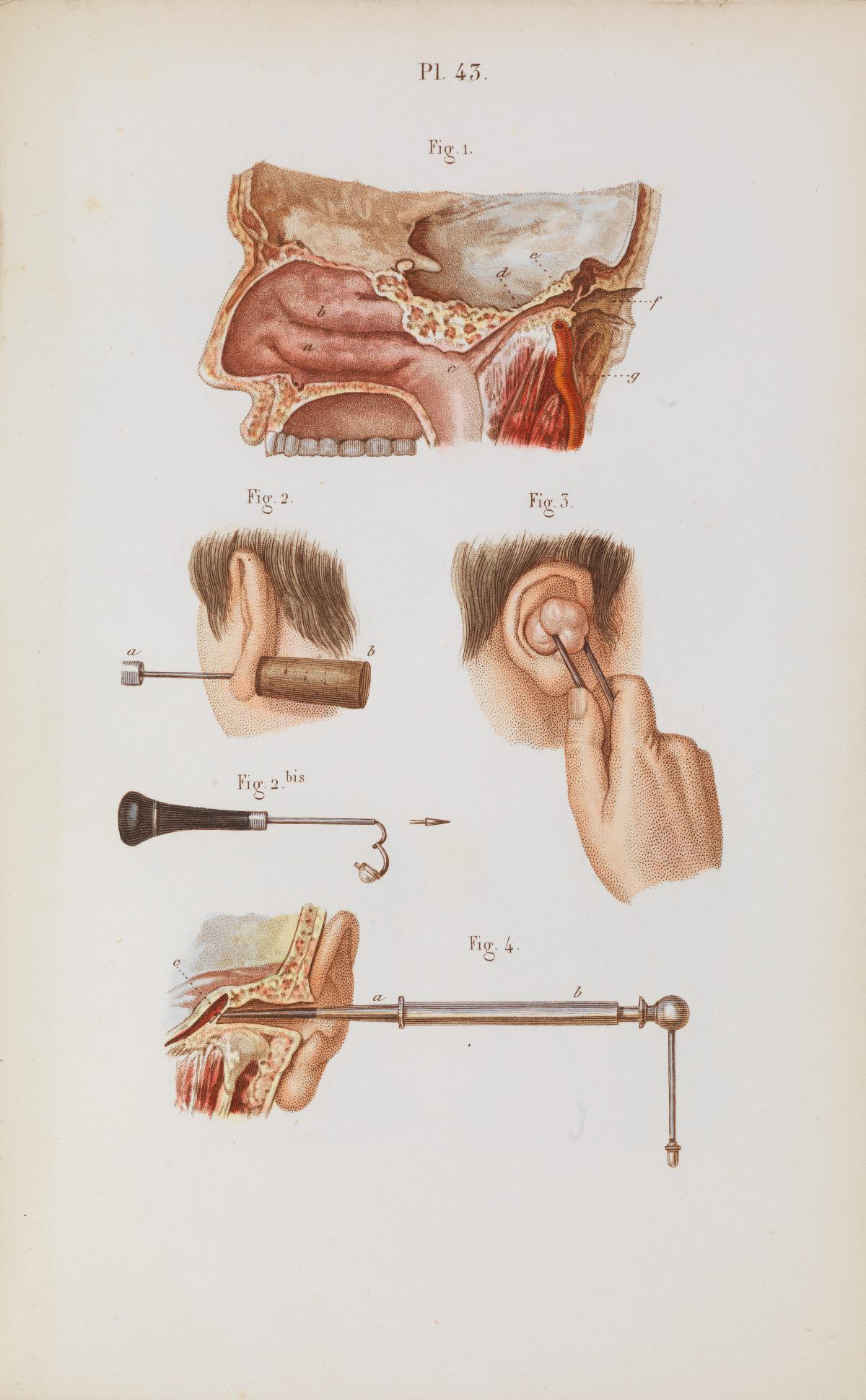
Plate 43 reads like a lesson in early ear surgery, combining medical illustration with the calm precision of a teaching chart. At the top, a labeled cross-section of the head traces the passage from outer ear toward the inner spaces of the skull, rendered in soft washes that make bone, tissue, and cavity easy to distinguish. The numbering of “Fig.” details and lettered markers signals a page meant to be studied, not merely admired.
Below, the focus shifts from anatomy to technique, showing the ear in close-up alongside specialized instruments. One figure demonstrates a tool aligned with the ear canal, while another depicts a practitioner’s hand using forceps-like implements, emphasizing careful placement and controlled movement. The separate instrument drawings—handles, shafts, and attachments—offer a catalog-like clarity, useful for understanding how historical surgical tools were designed to reach delicate structures.
For readers interested in the history of medicine, otology, or anatomical art, this plate provides a vivid snapshot of how surgeons and students once learned procedures through print. The crisp layout, restrained color, and systematic labeling make it especially searchable and relevant for topics like ear anatomy, surgical illustration, and vintage medical diagrams. As an artwork, it also reminds us how craft and science met on the page, turning complex bodily knowledge into a readable, teachable image.