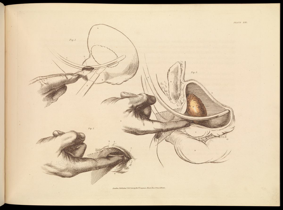
Plate XIX turns surgical instruction into a carefully staged narrative, using fine linework and labeled figures to guide the viewer through the removal of a bladder stone. The composition focuses on hands, instruments, and anatomy rather than faces, keeping attention on technique and the precise orientation of tissues. Seen as an artwork as much as a medical document, the print balances clinical clarity with the quiet drama of an operation rendered in ink.
Across the different figures, the illustrator isolates key moments: the approach of a tool, the positioning of the surgeon’s fingers, and a cutaway view that reveals the stone nestled within the bladder. Cross-hatching and subtle shading create depth, while the spare background mimics the clean, didactic style of early medical illustration. Even without a stated date or place on the page, the plate belongs to a tradition of anatomical teaching images designed for careful study and repeated reference.
For readers interested in the history of surgery, urology, and anatomical art, this historical plate offers a candid look at how complex procedures were explained before modern photography and imaging. The emphasis on step-by-step visualization hints at the apprenticeship culture of medicine, where knowledge traveled through printed atlases and shared diagrams. As a WordPress post feature, it invites reflection on the evolution of surgical practice, medical education, and the enduring power of illustration to make hidden anatomy visible.