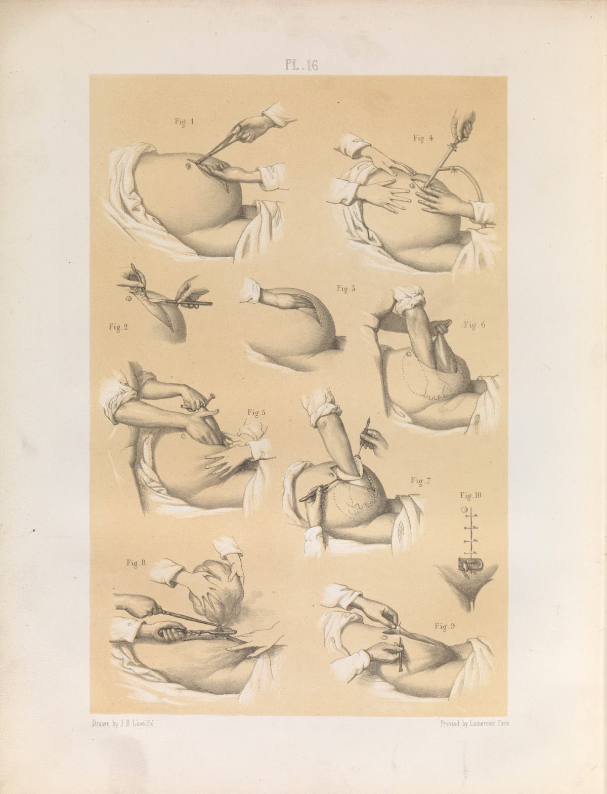
Plate 16 reads like a surgical lesson rendered in ink and wash, arranging numbered figures across a single sheet to guide the eye through “various operative stages” in removing a tumour from the uterus. Hands, instruments, and draped fabric take center stage while the patient’s identity is deliberately absent, a reminder that medical illustration often balanced clinical clarity with modesty. The calm, systematic composition signals an educational purpose, designed to be consulted and re-consulted rather than simply viewed.
Across the sequence, the illustrator focuses on technique: grasping and repositioning tissue, managing exposure, and working within a confined operative field. Several vignettes highlight the coordinated roles of multiple hands—one stabilizing, another manipulating a tool—suggesting the teamwork and choreography required in gynecological surgery. Even without narrative text on the page, the progression of figures implies a step-by-step approach meant for students and practitioners.
As an artwork, this plate also belongs to the broader history of medical publishing, where precise drawings functioned as technology in their own right, transmitting knowledge before photography became standard in the operating room. Its restrained palette and clean labeling make it especially searchable for readers interested in historical obstetrics and gynecology, surgical instruments, and anatomical illustration. For modern viewers, the sheet offers a candid window into how earlier generations tried to standardize complex procedures on paper—part science, part craft, and unmistakably of its time.