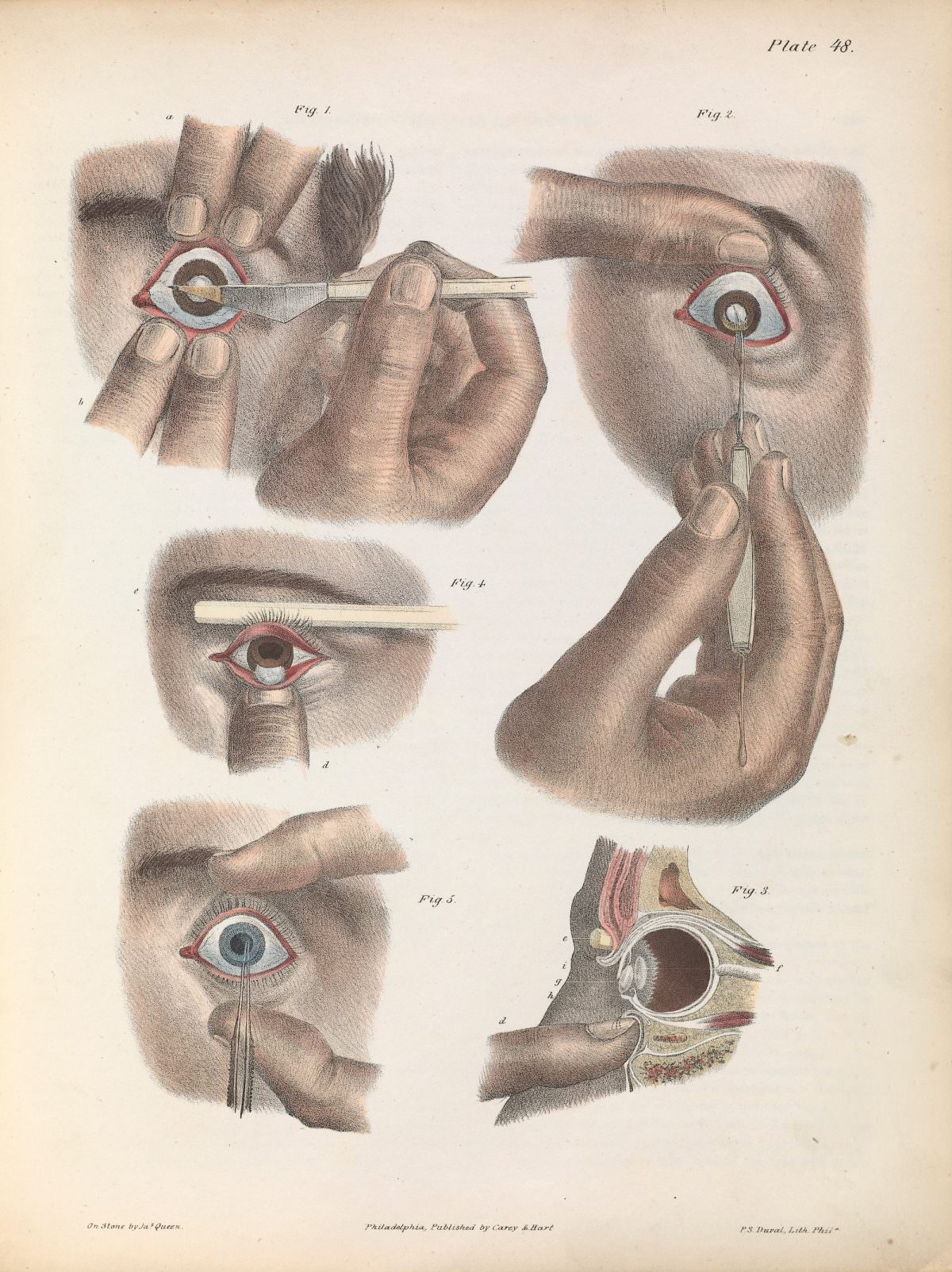
Plate 48 lays out cataract surgery as a carefully choreographed sequence, rendered with the calm precision of a medical atlas. Multiple figures guide the viewer through an “operation by extraction,” focusing on the inferior section of the cornea, with each hand position and instrument angle presented as if in slow motion. The restrained palette and crisp outlines turn a delicate procedure into something teachable—an anatomical lesson meant for study as much as for awe.
In the upper scenes, the eyelids are held open and the eye is stabilized while a blade approaches the lower corneal edge, emphasizing control and steadiness at every step. Elsewhere, slender tools and forceps are shown entering the eye with almost geometric clarity, while a cross-section diagram reveals the internal structures that make the operation possible. Small labels and figure numbers reinforce the instructional intent, inviting close reading from students of ophthalmology and the history of surgery.
Beyond its clinical purpose, the illustration also speaks to the era’s faith in specialized instruments, standardized technique, and printed knowledge shared across distances. For readers interested in historical medical illustration, cataract removal, and the development of eye surgery, this plate offers a vivid snapshot of how surgeons communicated complex procedures before modern photography and video. It remains a striking artifact at the intersection of art, anatomy, and the long pursuit of restored sight.