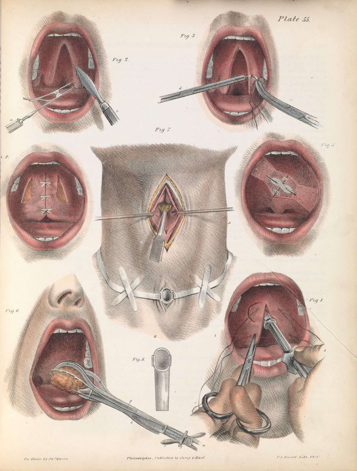
Plate 55 from J. Pancoast’s *A Treatise on Operative Surgery* (1846) reads like a carefully staged lesson in nineteenth-century technique, rendered with the calm precision of a medical atlas. Multiple views of an open mouth dominate the sheet, each labeled figure isolating a different step—retractors drawing back tissue, scissors and forceps poised at the throat, and sutures neatly marching across a repaired surface. The hand-colored tones of lips, palate, and tongue bring an unsettling immediacy, reminding modern viewers that these were practical guides for surgeons rather than mere “artworks.”
Across the composition, the illustrator’s goal is clarity: instruments are angled to reveal their working ends, and the anatomy is simplified just enough to make the operative field legible. Several scenes focus on the back of the mouth and soft palate, where stitches, clamps, and careful cutting suggest reconstructive procedures and the management of delicate structures. A central vignette shifts away from the face to a small, strapped operative window, reinforcing how surgical manuals taught control—of the patient’s body, the incision, and the surgeon’s hands—through staged diagrams.
For readers interested in the history of surgery, medical illustration, or nineteenth-century healthcare, this 1846 plate offers a vivid entry point into pre-modern operating practice as it was taught on the page. It also highlights the period’s reliance on detailed printed visuals to standardize technique, long before photography became routine in medical education. As a WordPress post feature, the image works well for discussions of antique medical prints, operative surgery textbooks, and the evolving relationship between art, anatomy, and clinical instruction.