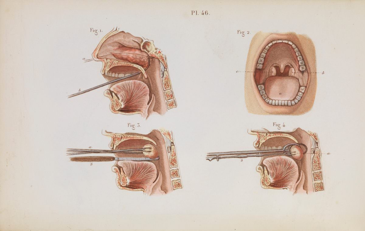
Plate 46 offers a meticulous medical illustration devoted to procedures of the upper airway, pairing the removal of nasal polyps with a tonsillectomy. Rendered as a teaching aid, the page is arranged into multiple labeled figures that guide the eye from the nasal cavity down toward the throat, emphasizing anatomy as much as technique. The calm, clinical presentation—lettered parts, clean margins, and carefully shaded tissue—reflects how surgical knowledge was codified for study and repetition.
In the cross-sectional views, slender instruments enter through the mouth to reach the back of the nasal passages, where growths could obstruct breathing and alter speech. One sequence suggests a step-by-step approach: positioning, grasping or looping, and extraction, each stage captured with precise linework. The open-mouth diagram complements these cutaway scenes by orienting the viewer to the tonsillar region and the surrounding structures a surgeon would need to avoid.
Such historical medical plates sit at the crossroads of art and practice, translating invasive operations into legible diagrams for students and practitioners. For readers interested in the history of otolaryngology (ENT), surgical instruments, and medical education, this artwork preserves the visual language of an earlier operating room—where anatomy was learned as much from printed color plates as from the body itself. Its enduring value lies in that blend of instruction and craftsmanship, making Plate 46 a compelling artifact for both medical history and illustration enthusiasts.