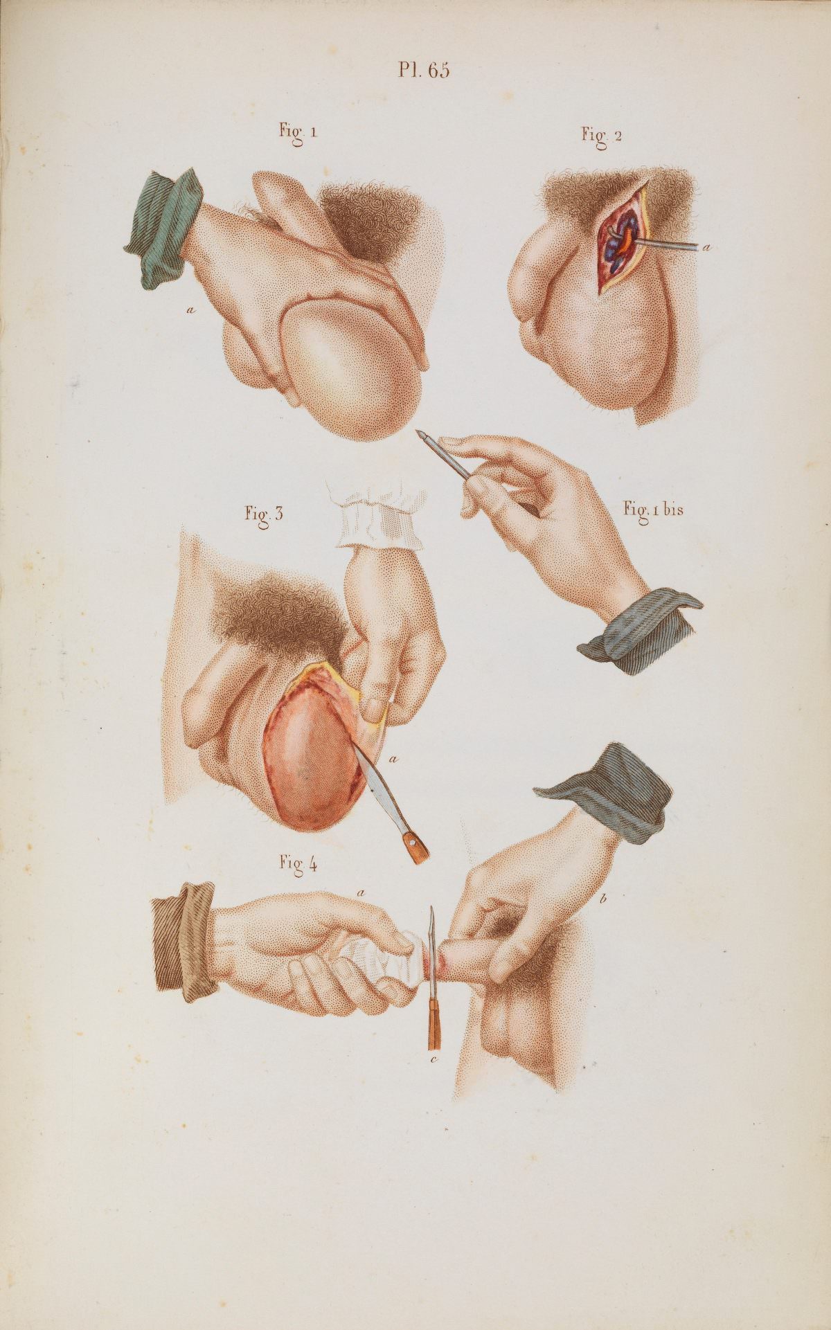
Plate 65 reads like a page torn from a surgeon’s notebook, rendered with the calm precision of medical art. Numbered figures map the anatomy of the scrotum and the careful placement of hands and instruments, turning an intimate and difficult subject into something instructional. The restrained palette, fine stippling, and clean margins suggest an illustration meant to be studied closely, not merely glanced at.
Across the separate panels, the procedure is broken into stages: the area prepared and supported, an incision opened to reveal layered tissue, and the tumour approached with deliberate cuts. A standalone hand holding a scalpel underscores technique as much as anatomy, while lettered points guide the viewer’s eye as if following a lesson. Despite the clinical intent, the illustrator’s attention to skin, hair, and light gives the plate an unmistakably human immediacy.
For historians of medicine, this artwork offers a window into how surgical knowledge was shared before photography dominated textbooks—through didactic, carefully composed plates that balanced clarity with realism. It also speaks to the evolution of urological and general surgery, where successful outcomes depended on anatomy, steadiness, and the ability to communicate method on the printed page. As a historical medical illustration, “Surgical removal of tumours from the scrotum” remains a striking example of how art and science met in the service of teaching.