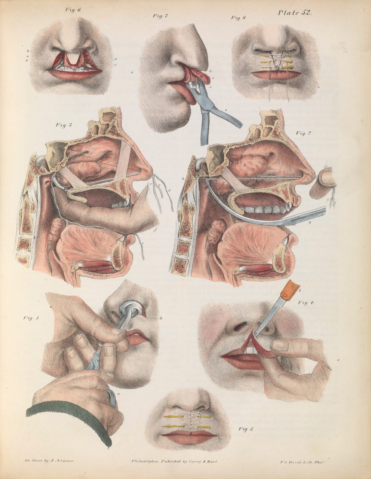
Across this tightly arranged medical plate, a sequence of numbered figures guides the eye through procedures of removal and repair, rendered with the calm precision of a surgical textbook. Forceps and a small knife appear in close-up alongside careful views of the lips, while cross-sections of the mouth and nasal cavity reveal the hidden anatomy behind each step. The palette is restrained but purposeful, using color to emphasize tissue, incisions, and the boundaries surgeons needed to understand.
In the upper figures, “torsion and traction” with forceps and the alternative method “by ligature” read like a practical comparison of techniques, meant to be studied rather than merely observed. The composition balances instruments, hands, and patient profiles to demonstrate exactly how traction is applied and where ligatures are placed. Even without naming a specific practitioner, the plate conveys an era when surgical knowledge was taught through engraved or lithographed illustrations that could travel farther than any lecture.
Attention then turns to “simple hare-lip” and the more complex “double” and “complicated” forms, with sutures shown bridging the split and holding tissue in alignment. These diagrams underline how reconstructive surgery depended on both anatomical insight and methodical planning, from incision lines to final closure. For readers interested in the history of surgery, cleft lip repair, or antique medical illustration, this artwork offers a vivid window into how clinicians once learned to operate—one labeled figure at a time.